Supreme Court shown 4D ultrasound images, urged to uphold Mississippi 15-week abortion ban

A nonprofit organization has submitted an amicus brief to the U.S. Supreme Court featuring ultrasound imagery over time to demonstrate how scientific advancements justify overturning the long-standing precedent in American abortion jurisprudence.
As the U.S. Supreme Court is slated to hear a major challenge to Roe v. Wade, a lawyer writing on behalf of the Catholic Association Foundation and three medical doctors filed the brief in support of the state of Mississippi in the case of Dobbs v. Jackson Women’s Health Organization last month.
In this case, billed by pro-life advocates as a “landmark opportunity” to chip away at the 1973 Roe v. Wade decision that legalized abortion nationwide, the state of Mississippi is asking the justices to reverse a lower court decision finding that its 15-week abortion ban is unconstitutional.
The Catholic Association Foundation was “granted special permission from the Supreme Court” to include ultrasound imagery as part of its amicus brief, according to information obtained by The Christian Post.
The brief noted that when striking down Mississippi’s ban on abortions after 15 weeks gestation, the U.S. Fifth Circuit Court of Appeals cited the 1992 Supreme Court decision Planned Parenthood v. Casey, which determined that “[n]o state interest is constitutionally adequate to ban abortions before viability.”
The term “viability” refers to a fetus’ ability to survive outside the womb. In 1973, when Roe was decided, 28 weeks was seen as the point of viability. By 1992, the term “viable” applied to babies born at 23 or 24 weeks gestation.
“Roe and Casey’s viability standard is incomplete and outdated according to current science,” the brief declared.
The counsel for the amici explained that babies born at 21 weeks gestation are now capable of surviving outside the womb.
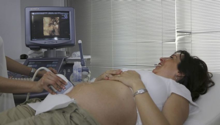
Additionally, the brief lamented that “the human form of the child, regardless of its viability, is unaccounted for by Casey.” It pointed to ultrasound technology as a source of a “clear window into the womb to witness the humanity of the unborn child.”
The brief included ultrasound images from the 1970s and the 1980s and modern ultrasound images incorporating the use of three-dimensional and four-dimensional technology.
“Sonograms in the 1970s were rudimentary,” the document argued.
While “sonograms in the late 1980s were still blurry and indefinite,” the technology has since improved substantially. A caption accompanying one set of modern ultrasound images detailed how “3D and 4D images, surface rendered, reveal the human face of the fetus: plump cheeks, delicately formed lips, and tiny noses.”
“In 4D renderings, we can observe movement. The baby can be seen yawning, sucking her thumb, and kicking," the brief continued. "4D technology also shows fleeting expressions: the grimace of a cry, a frown wrinkling the forehead, a curve of the lips into a smile. The baby’s mouth opens, and we can even see the tongue moving. These detailed images have opened a window into the womb, allowing us to witness the human form of the child."
The brief contends that unborn babies at 15 weeks gestation look “unmistakably human,” and the state has an interest in protecting them.
"At 15 weeks gestation, all major organs are formed and functioning, including the liver, kidneys, pancreas, and brain,” the brief reads.
“Although the child receives nutrients and oxygen through the umbilical cord, the digestive, urinary, and respiratory systems are practicing for extra-uterine life. At 15 weeks, the fetus swallows and urinates; she even ‘breathes,’ filling her lungs with amniotic fluid and expelling it. The cardiovascular system is fully formed. Not only is the baby’s heartbeat detectable, as it has been for nine weeks, but the four chambers of the fetal heart are visible."
In addition to highlighting a clear fetal profile consisting of a “gently sloping nose ... distinct upper and lower lips and chin,” one ultrasound image featured in the brief is credited with making “external genitalia” visible. This enables “sonographers to inform families whether their baby is a son or daughter.” Although ultrasound images account for most of the pictures in the brief, the document also includes photographs of fetal surgeries to emphasize that modern medicine now views the fetus as “a patient in her own right.”
The brief concluded that “the Court should relinquish its role as nationwide abortion regulator and return the job to the States and elected officials where it belongs.” The court is expected to hear oral arguments in Dobbs v. Jackson Women’s Health Organization in its upcoming October term. A decision is expected by next spring.
Ryan Foley is a reporter for The Christian Post. He can be reached at: ryan.foley@christianpost.com


